Pelvic Anatomy - Nerves Of Male Pelvis Overview Preview Human Anatomy Kenhub Youtube. The pelvic girdle and pelvic spine. Use the mouse scroll wheel to move the images up and down alternatively use the tiny arrows (>>) on both side of the image to move the images.>>) on both side of the image to move the images. The bony pelvis consists of the two hip bones (also known as innominate or pelvic bones), the sacrum and the coccyx. However, knowledge of the anatomy of various structures that surround these organs has evolved over time. The right and left hip bones also converge anteriorly to attach to each other.
Understanding what each term refers to and its purpose can help you make the wisest decision. The video is narrated by one of our awesome anatomists, dr. S1 supplies the lateral aspect of the sole. Reproduction system pelvis female woman reproductive system pelvic floor women female bladder and urethra female pelvic floor pelvis woman pelvic floor health woman incontinence pelvis muscles. Regarding the surface anatomy, the perineal area is the region between the thighs, extending from the pubic symphysis anteriorly to the gluteal folds posteriorly.
Pelvic Anatomy Muscoloskeletal Portfolio from sites.google.com Gross anatomy of the pelvis—namely the bladder, uterus, fallopian tubes, ovaries, rectum, and the muscles—has remained unchanged; Laparoscopic anatomy of the female pelvic region. The anatomy of the pelvis varies depending on whether you are male or female. Surgical anatomy of the female pelvis by laparoscopy. Understanding what each term refers to and its purpose can help you make the wisest decision. The lining of the uterus. It provides attachment to some important muscles in the region, and forms a cavity which accommodates several important internal organs. It is inferior to the pelvic diaphragm.
Classic anatomical studies have provided few details of the inferior hypogastric plexus morphology or the location and nature of the associated nerves.
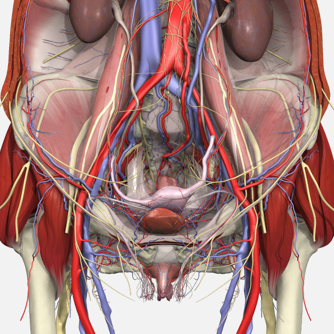
The male pelvis is different from a female's. The inferior hypogastric plexus is also known as the pelvic ganglion. The fusion of the pelvic splanchnic nerves, sacral splanchnic nerves, and superior hypogastric plexus along with visceral afferent fibers forms the inferior hypogastric plexus. By continuing to browse this site you are agreeing to our use of cookies. It is located in the middle of the pelvis between the urinary bladder lying before and the large bowel lying behind it. Surgical pelvic anatomy in gynecologic oncology shailesh puntambekar1,* | ranjit manchanda2 this is an open access article under the terms of the creative commons attribution license, which permits use, distribution and reproduction in any medium, provided the original work is properly cited. However, knowledge of the anatomy of various structures that surround these organs has evolved over time. Pelvic anatomy www.freelivedoctor.com slideshare uses cookies to improve functionality and performance, and to provide you with relevant advertising. It is inferior to the pelvic diaphragm. The pelvic girdle (hip girdle) is formed by a single bone, the hip bone or coxal bone (coxal = hip), which serves as the attachment point for each lower limb. Anatomy of female pelvic area facebook twitter linkedin pinterest print fertility and reproductive health pelvic floor disorders fertility, pregnancy and childbirth women's health. See female pelvic anatomy stock video clips. Understanding what each term refers to and its purpose can help you make the wisest decision.
This video provides an overview of pelvic floor anatomy including key muscles and their functions. Regarding the surface anatomy, the perineal area is the region between the thighs, extending from the pubic symphysis anteriorly to the gluteal folds posteriorly. There are four articulations within the pelvis: The pelvic floor muscles form part of the pelvic floor and play a critical role in sexual function as well as the maintenance of urinary and faecal continence, anatomy of the prostate gland edit source The uterus represents the essential landmark of pelvic anatomy.
Pelvic Floor Disorders Anatomy Primal Pictures from www.primalpictures.com It's located between the abdomen and the legs. On a sagittal plane, the uterus has a pyriform shape: The lining of the uterus. The pelvis's frame is made up of the bones of the pelvis, which connect the axial skeleton to the femurs, and therefore acts in weight bearing of the upper body. Ct body (lymph nodes) ct. It provides attachment to some important muscles in the region, and forms a cavity which accommodates several important internal organs. The anatomy of the pelvis varies depending on whether you are male or female. It is usually divided into two separate anatomic regions:
Ct body (lymph nodes) ct.
The bony pelvis consists of the two hip bones (also known as innominate or pelvic bones), the sacrum and the coccyx. The pelvic bones are smaller and narrower. The uterus represents the essential landmark of pelvic anatomy. Clinical anatomy the lumbosacral trunk (l4, l5) and the ventral ramus of nerve s1 cross the nerves of the pelvis surface of the joint and may be involved in the disease of the joints, causing pain in the area of their distribution below the knee. The pelvis (plural pelves or pelvises) is either the lower part of the trunk of the human body between the abdomen and the thighs (sometimes also called pelvic region of the trunk) or the skeleton embedded in it (sometimes also called bony pelvis, or pelvic skeleton). {{configctrl2.info.metadescription}} this site uses cookies. L4 supplies the medial aspect of leg and sole. This video provides an overview of pelvic floor anatomy including key muscles and their functions. There are four articulations within the pelvis: This area provides support for the intestines and also contains the bladder and reproductive organs. The anatomy of the pelvis varies depending on whether you are male or female. Anatomy of female pelvic area facebook twitter linkedin pinterest print fertility and reproductive health pelvic floor disorders fertility, pregnancy and childbirth women's health. Ct body (lymph nodes) ct.
The uterus, tubes and ovaries are attached to the woman with a number of structures, many of them misnamed. It provides attachment to some important muscles in the region, and forms a cavity which accommodates several important internal organs. The pelvic girdle (hip girdle) is formed by a single bone, the hip bone or coxal bone (coxal = hip), which serves as the attachment point for each lower limb. However, knowledge of the anatomy of various structures that surround these organs has evolved over time. Ct body (lymph nodes) ct.
Anatomy Of The Pelvic Cavity Osmosis from d16qt3wv6xm098.cloudfront.net It is inferior to the pelvic diaphragm. Gross anatomy of the pelvis—namely the bladder, uterus, fallopian tubes, ovaries, rectum, and the muscles—has remained unchanged; Each hip bone, in turn, is firmly joined to the axial skeleton via its attachment to the sacrum of the vertebral column. Ct body (lymph nodes) ct. The uterus, sometimes called the womb, is a muscular organ located in the pelvis. The pelvis is a basin shaped bony structure formed by the combination of two pelvic bones (hip bones or innominate bones) and the sacrum. The anatomy of the pelvis varies depending on whether you are male or female. The pelvis is the lower part of the torso.
Reproduction system pelvis female woman reproductive system pelvic floor women female bladder and urethra female pelvic floor pelvis woman pelvic floor health woman incontinence pelvis muscles.
Each hip bone, in turn, is firmly joined to the axial skeleton via its attachment to the sacrum of the vertebral column. Gross anatomy of the pelvis—namely the bladder, uterus, fallopian tubes, ovaries, rectum, and the muscles—has remained unchanged; Reproduction system pelvis female woman reproductive system pelvic floor women female bladder and urethra female pelvic floor pelvis woman pelvic floor health woman incontinence pelvis muscles. Understanding what each term refers to and its purpose can help you make the wisest decision. The pelvic girdle and pelvic spine. The pelvis is a basin shaped bony structure formed by the combination of two pelvic bones (hip bones or innominate bones) and the sacrum. The broad ligament is not really a ligament but rather two leaves of peritoneum in between which run the uterine vessels, lymphatics and nerves. This area provides support for the intestines and also contains the bladder and reproductive organs. The uterus, sometimes called the womb, is a muscular organ located in the pelvis. Pelvic anatomy www.freelivedoctor.com slideshare uses cookies to improve functionality and performance, and to provide you with relevant advertising. The fusion of the pelvic splanchnic nerves, sacral splanchnic nerves, and superior hypogastric plexus along with visceral afferent fibers forms the inferior hypogastric plexus. The video is narrated by one of our awesome anatomists, dr. Anatomy of female pelvic area facebook twitter linkedin pinterest print fertility and reproductive health pelvic floor disorders fertility, pregnancy and childbirth women's health.
Share :
Post a Comment
for "Pelvic Anatomy - Nerves Of Male Pelvis Overview Preview Human Anatomy Kenhub Youtube"


Post a Comment for "Pelvic Anatomy - Nerves Of Male Pelvis Overview Preview Human Anatomy Kenhub Youtube"